 Login / Register


- Clone
- YN1/1.7.4 (See other available formats)
- Regulatory Status
- RUO
- Other Names
- ICAM-1, Ly-47
- Isotype
- Rat IgG2b, κ
- Ave. Rating
- Submit a Review
- Product Citations
- publications

-

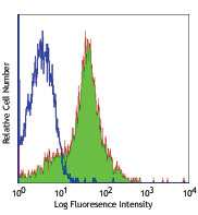
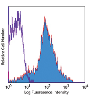
C57BL/6 mouse splenocytes stained with biotinylated YN1/1.7.4 , followed by Sav-PE
| Cat # | Size | Price | Quantity Check Availability | Save | ||
|---|---|---|---|---|---|---|
| 116103 | 50 µg | £57 | ||||
CD54 is a 90 kD immunoglobulin superfamily member also known as ICAM-1 and Ly-47. It is expressed on activated endothelial cells, high endothelial venules (HEV), T and B cells, monocytes/ macrophages, granulocytes, and dendritic cells. CD54 is an important intracellular adhesion molecule that participates in T cell-T cell, T cell-B cell, and T cell-target cell interactions via binding of LFA-1 (CD11a/CD18) and Mac-1 (CD11b/CD18). CD54 has also been shown to be involved in lymphocyte trafficking, making it an important molecule in many immune reactions and inflammation. CD54 is also a receptor for rhinovirus. The YN1/1.7.4 antibody has been reported to block binding of mouse CD54 to LFA-1 and Mac-1, inhibit cell-cell adhesion, and function in antigen presentation to T cells and leukocyte migration to inflammatory tissues.
Product DetailsProduct Details
- Verified Reactivity
- Mouse
- Antibody Type
- Monoclonal
- Host Species
- Rat
- Immunogen
- Mouse NS-1 cells
- Formulation
- Phosphate-buffered solution, pH 7.2, containing 0.09% sodium azide.
- Preparation
- The antibody was purified by affinity chromatography, and conjugated with biotin under optimal conditions.
- Concentration
- 0.5 mg/ml
- Storage & Handling
- The antibody solution should be stored undiluted between 2°C and 8°C. Do not freeze.
- Application
-
FC - Quality tested
- Recommended Usage
-
Each lot of this antibody is quality control tested by immunofluorescent staining with flow cytometric analysis. For flow cytometric staining, the suggested use of this reagent is ≤ 0.25 µg per 106 cells in 100 µl volume. It is recommended that the reagent be titrated for optimal performance for each application.
- Application Notes
-
Additional reported applications (for the relevant formats) include: in vitro and in vivo blocking of cell-cell adhesion and CD54 functions1,2,4, immunohistochemical staining3 of acetone-fixed frozen sections, immunoprecipitation4, and Western blotting (non-reducing). The Ultra-LEAF™ purified antibody (Endotoxin < 0.01 EU/µg, Azide-Free, 0.2 µm filtered) is recommended for functional assays (Cat. Nos. 116131-116136).
-
Application References
(PubMed link indicates BioLegend citation) -
- Kumaska T, et al. 1996. J. Clin. Invest. 97:2362. (Block)
- Horley KJ, et al. 1989. EMBO J. 8:2889. (Block)
- Burns AR, et al. 1994. J. Immunol. 153:3189. (IHC)
- Takei F, et al. 1985. J. Immunol. 134:1403. (Block, IP)
- Sumagin R, et al. 2010. J. Immunol. 184:5242. PubMed
- Bankoti J, et al. 2010. Toxicol. Sci. 115:422. (FC) PubMed
- Seidler DG, et al. 2011 J. Immunol. 187:6108. PubMed
- Chacko AM, et al. 2011. Curr Opin Colloid Interface Sci. 16:215. PUbMed
- El-Assaad F, et al. 2013. Infect Immun. 81:3984. PubMed
- Greineder CF, et al. 2013. PLoS One. 14:80110. PubMed
- Product Citations
-
- RRID
-
AB_313694 (BioLegend Cat. No. 116103)
Antigen Details
- Structure
- Ig superfamily, 90 kD
- Distribution
-
Activated endothelial cells, high endothelial venules, T cells and B cells, monocytes/macrophages, granulocytes, dendritic cells
- Function
- Immune reaction, inflammation, adhesion
- Ligand/Receptor
- CD11a/CD18 (LFA-1) or CD11b/CD18 (Mac-1) and CD11c/CD18, CD43, hyaluronan, fibrinogen
- Cell Type
- B cells, Dendritic cells, Endothelial cells, Granulocytes, Macrophages, Mesenchymal Stem Cells, Monocytes, T cells
- Biology Area
- Cell Adhesion, Cell Biology, Costimulatory Molecules, Immunology, Innate Immunity, Neuroscience, Neuroscience Cell Markers, Stem Cells
- Molecular Family
- Adhesion Molecules, CD Molecules
- Antigen References
-
1. Barclay AN, et al. 1997. The Leukocyte Antigen FactsBook Academic Press.
2. Springer TA. 1990. Nature 346:425.
3. Rothlein R, et al. 1986. J. Immunol. 137:1270. - Gene ID
- 15894 View all products for this Gene ID
- UniProt
- View information about CD54 on UniProt.org
Related Pages & Pathways
Pathways


Related FAQs
- How many biotin molecules are per antibody structure?
- We don't routinely measure the number of biotins with our antibody products but the number of biotin molecules range from 3-6 molecules per antibody.
Other Formats
View All CD54 Reagents Request Custom Conjugation| Description | Clone | Applications |
|---|---|---|
| Purified anti-mouse CD54 | YN1/1.7.4 | FC,IHC-F,IP,WB |
| Biotin anti-mouse CD54 | YN1/1.7.4 | FC |
| FITC anti-mouse CD54 | YN1/1.7.4 | FC |
| PE anti-mouse CD54 | YN1/1.7.4 | FC |
| Alexa Fluor® 488 anti-mouse CD54 | YN1/1.7.4 | FC |
| Alexa Fluor® 647 anti-mouse CD54 | YN1/1.7.4 | FC |
| Pacific Blue™ anti-mouse CD54 | YN1/1.7.4 | FC |
| APC anti-mouse CD54 | YN1/1.7.4 | FC |
| PerCP/Cyanine5.5 anti-mouse CD54 | YN1/1.7.4 | FC |
| PE/Cyanine7 anti-mouse CD54 | YN1/1.7.4 | FC |
| APC/Fire™ 750 anti-mouse CD54 | YN1/1.7.4 | FC |
| TotalSeq™-A0074 anti-mouse CD54 | YN1/1.7.4 | PG |
| PE/Dazzle™ 594 anti-mouse CD54 | YN1/1.7.4 | FC |
| Ultra-LEAF™ Purified anti-mouse CD54 | YN1/1.7.4 | FC,IP,WB |
| TotalSeq™-C0074 anti-mouse CD54 | YN1/1.7.4 | PG |
| TotalSeq™-B0074 anti-mouse CD54 | YN1/1.7.4 | PG |
| Brilliant Violet 421™ anti-mouse CD54 | YN1/1.7.4 | FC |
| Brilliant Violet 711™ anti-mouse CD54 | YN1/1.7.4 | FC |
| Brilliant Violet 785™ anti-mouse CD54 | YN1/1.7.4 | FC |
| Brilliant Violet 605™ anti-mouse CD54 | YN1/1.7.4 | FC |
Customers Also Purchased
Compare Data Across All Formats
This data display is provided for general comparisons between formats.
Your actual data may vary due to variations in samples, target cells, instruments and their settings, staining conditions, and other factors.
If you need assistance with selecting the best format contact our expert technical support team.
-
Purified anti-mouse CD54

C57BL/6 mouse splenocytes stained with purifid YN1/1.7.4 , f... 
C57BL/6 mouse frozen testis section was fixed with 4% parafo... -
Biotin anti-mouse CD54

C57BL/6 mouse splenocytes stained with biotinylated YN1/1.7.... -
FITC anti-mouse CD54

C57BL/6 splenocytes stained with YN1/1.7.4 FITC -
PE anti-mouse CD54

C57BL/6 mouse splenocytes stained with YN1/1.7.4 PE -
Alexa Fluor® 488 anti-mouse CD54

C57BL/6 splenocytes stained with YN1/1.7.4 Alexa Fluor® 488 -
Alexa Fluor® 647 anti-mouse CD54

C57BL/6 mouse splenocytes stained with YN1/1.7.4 Alexa Fluor... -
Pacific Blue™ anti-mouse CD54

C57BL/6 mouse splenocytes stained with YN1/1.7.4 Pacific Blu... -
APC anti-mouse CD54

C57BL/6 splenocytes stained with YN1/1.7 APC -
PerCP/Cyanine5.5 anti-mouse CD54

C57BL/6 mouse splenocytes were stained with CD54 (clone YN1/... -
PE/Cyanine7 anti-mouse CD54

C57BL/6 mouse splenocytes were stained with CD54 (clone YN1/... -
APC/Fire™ 750 anti-mouse CD54

C57BL/6 mouse splenocytes were stained with CD54 (clone YN1/... -
TotalSeq™-A0074 anti-mouse CD54
-
PE/Dazzle™ 594 anti-mouse CD54

C57BL/6 mouse splenocytes stained with CD54 (clone YN1/1.7.4... -
Ultra-LEAF™ Purified anti-mouse CD54
C57BL/6 mouse splenocytes stained with purifid YN1/1.7.4, fo... -
TotalSeq™-C0074 anti-mouse CD54
-
TotalSeq™-B0074 anti-mouse CD54
-
Brilliant Violet 421™ anti-mouse CD54

C57BL/6 mouse splenocytes stained with anti-mouse CD54 (clon... -
Brilliant Violet 711™ anti-mouse CD54

C57BL/6 splenocytes were stained with anti-mouse CD54 (clone... -
Brilliant Violet 785™ anti-mouse CD54

C57BL/6 mouse splenocytes were stained with anti-mouse CD54 ... -
Brilliant Violet 605™ anti-mouse CD54

C57BL/6 mouse splenocytes were stained with anti-mouse CD54 ...


Follow Us