 Login / Register


- Clone
- B27 (See other available formats)
- Regulatory Status
- RUO
- Other Names
- Interferon-γ, Immune interferon, Type II interferon, T cell interferon, Macrophage-activating factor (MAF), IFN-g, IFN-gamma
- Isotype
- Mouse IgG1, κ
- Ave. Rating
- Submit a Review
- Product Citations
- publications

-

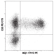
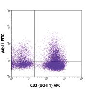
PMA+ionomycin-stimulated (6 hours) human peripheral blood lymphocytes intracellularly stained with B27 APC and CD3 (HIT3a) PE/Cy5
| Cat # | Size | Price | Quantity Check Availability | Save | ||
|---|---|---|---|---|---|---|
| 506510 | 100 tests | £228 | ||||
Interferon-γ is a potent multifunctional cytokine which is secreted primarily by activated NK cells and T cells. Originally characterized based on anti-viral activities, IFN-γ also exerts anti-proliferative, immunoregulatory, and proinflammatory activities. IFN-γ can upregulate MHC class I and II antigen expression by antigen-presenting cells. The B27 antibody reacts with the human interferon-γ. The B27 antibody can neutralize the bioactivity of natural or recombinant IFN-γ.
Product DetailsProduct Details
- Verified Reactivity
- Human
- Reported Reactivity
- Chimpanzee, Baboon, Cynomolgus, Rhesus, African Green, Pigtailed Macaque, Sooty Mangabey
- Antibody Type
- Monoclonal
- Host Species
- Mouse
- Immunogen
- E. coli-expressed recombinant human IFN-γ
- Formulation
- Phosphate-buffered solution, pH 7.2, containing 0.09% sodium azide and BSA (origin USA)
- Preparation
- The antibody was purified by affinity chromatography, and conjugated with APC under optimal conditions.
- Storage & Handling
- The antibody solution should be stored undiluted between 2°C and 8°C, and protected from prolonged exposure to light. Do not freeze.
- Application
-
ICFC - Quality tested
- Recommended Usage
-
Each lot of this antibody is quality control tested by intracellular immunofluorescent staining with flow cytometric analysis. For flow cytometric staining, the suggested use of this reagent is 5 µl per million cells in 100 µl staining volume or 5 µl per 100 µl of whole blood.
- Excitation Laser
-
Red Laser (633 nm)
- Application Notes
-
Flow Cytometry2: The fluorochrome-labeled B27 antibody is useful for intracellular immunofluorescent staining and flow cytometric analysis to identify IFN-? -producing cells within mixed cell populations. For intracellular cytokine staining protocol, please visit www.biolegend.com and click on the support section.
Neutralization1,3,6,7: The Ultra-LEAF™ Purified antibody (Endotoxin <0.01 EU/µg, Azide-Free, 0.2 µm filtered) is recommended for neutralization of human IFN-? bioactivity (Cat. No. 506531). -
Application References
(PubMed link indicates BioLegend citation) -
- Favre C, et al. 1989. Molec. Immunol. 26:17. (Neut)
- Kaur A, et al. 2002. J Virol. 76:3646.
- Abrams J, et al. 1992. Immunol. Rev. 127:5. (Neut)
- Andersson U, et al. 1999. Detection and quantification of gene expression. New York:Springer-Verlag.
- Rout N, et al. 2010. PLoS One 5:e9787. (FC)
- Acosta-Rodriguez EV, et al. 2007. Nat. Immunol. 9:942-9. (Neut)
- Gangur V, et al. 1998. FASEB J. 12:705-13. (Neut)
- Product Citations
-
- RRID
-
AB_315443 (BioLegend Cat. No. 506510)
Antigen Details
- Structure
- Cytokine; dimer; 20-25 kD (Mammalian)
- Bioactivity
- Antiviral/antiparasitic activities; inhibits proliferation; enhances MHC class I and II expression on APC
- Cell Sources
- CD8+ and CD4+ T cells, NK cells
- Cell Targets
- T cells, B cells, macrophages, NK cells, endothelial cells, fibroblasts
- Receptors
- IFN-γRα (CDw119) dimerized with IFN-γRβ (AF-1)
- Cell Type
- Tregs
- Biology Area
- Cell Biology, Immunology, Neuroinflammation, Neuroscience
- Molecular Family
- Cytokines/Chemokines
- Antigen References
-
1. Fitzgerald K, et al. Eds. 2001. The Cytokine FactsBook. Academic Press San Diego.
2. De Maeyer E, et al. 1992. Curr. Opin. Immunol. 4:321.
3. Farrar M, et al. 1993. Annu. Rev. Immunol. 11:571.
4. Gray P, et al. 1987. Lymphokines 13:151. - Regulation
- Upregulated by IL-2, FGF-basic, EGF; downregulated by vitamin D3 or DMN; labile at pH2
- Gene ID
- 3458 View all products for this Gene ID
- UniProt
- View information about IFN-gamma on UniProt.org
Related Pages & Pathways
Pathways


Related FAQs
Other Formats
View All IFN-γ Reagents Request Custom Conjugation| Description | Clone | Applications |
|---|---|---|
| APC anti-human IFN-γ | B27 | ICFC |
| FITC anti-human IFN-γ | B27 | ICFC |
| PE anti-human IFN-γ | B27 | ICFC |
| Purified anti-human IFN-γ | B27 | ICFC,Neut,FC |
| Alexa Fluor® 700 anti-human IFN-γ | B27 | ICFC |
| PE/Cyanine7 anti-human IFN-γ | B27 | ICFC |
| Purified anti-human IFN-γ (Maxpar® Ready) | B27 | ICFC,CyTOF® |
| APC/Cyanine7 anti-human IFN-γ | B27 | ICFC |
| Pacific Blue™ anti-human IFN-γ | B27 | ICFC |
| PerCP/Cyanine5.5 anti-human IFN-γ | B27 | ICFC |
| PE/Dazzle™ 594 anti-human IFN-γ | B27 | ICFC |
| Ultra-LEAF™ Purified anti-human IFN-γ | B27 | ICFC,Neut,FC |
| Brilliant Violet 605™ anti-human IFN-γ | B27 | ICFC |
| Brilliant Violet 510™ anti-human IFN-γ | B27 | ICFC |
| Brilliant Violet 421™ anti-human IFN-γ | B27 | ICFC |
| TotalSeq™-B1003 anti-human IFN-γ | B27 | ICPG |
Customers Also Purchased
Compare Data Across All Formats
This data display is provided for general comparisons between formats.
Your actual data may vary due to variations in samples, target cells, instruments and their settings, staining conditions, and other factors.
If you need assistance with selecting the best format contact our expert technical support team.
-
APC anti-human IFN-γ

PMA+ionomycin-stimulated (6 hours) human peripheral blood ly... -
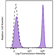
FITC anti-human IFN-γ

PMA+ionomycin-stimulated (6 hours) human peripheral blood ly... -
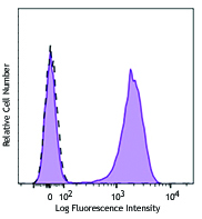
PE anti-human IFN-γ

PMA+ionomycin-stimulated (6 hours) human peripheral blood ly... -
Purified anti-human IFN-γ

PMA+ionomycin-stimulated human peripheral blood lymphocytes ... -
Alexa Fluor® 700 anti-human IFN-γ

PMA+ionomycin-stimulated (5 hours) human PBMCs surface stain... -
PE/Cyanine7 anti-human IFN-γ

PMA/ionomycin-stimulated (5 hours) human peripheral blood ly... -
Purified anti-human IFN-γ (Maxpar® Ready)

Human PBMCs were incubated for 6 hours in media alone (top) ... 
-
APC/Cyanine7 anti-human IFN-γ

PMA+ionomycin-stimulated (in the presence of monensin, 6hrs)... -
Pacific Blue™ anti-human IFN-γ

PMA+ionomycin-stimulated (in the presence of monensin, 6hrs)... 
-
PerCP/Cyanine5.5 anti-human IFN-γ

PMA+ionomycin-stimulated human peripheral blood lymphocytes... -
PE/Dazzle™ 594 anti-human IFN-γ

PMA+ionomycin-stimulated (in the presence of monensin, 6hrs)... 
-
Ultra-LEAF™ Purified anti-human IFN-γ
PMA+ionomycin-stimulated human peripheral blood lymphocytes ... -
Brilliant Violet 605™ anti-human IFN-γ

PMA+Ionomycin-stimulated (4 hours) human peripheral blood ly... -
Brilliant Violet 510™ anti-human IFN-γ

PMA+Ionomycin-stimulated (4 hours) human peripheral blood ly... -
Brilliant Violet 421™ anti-human IFN-γ

PMA+Ionomycin-stimulated (4 hours) human peripheral blood ly... -
TotalSeq™-B1003 anti-human IFN-γ


Follow Us