 Login / Register


- Clone
- AY13 (See other available formats)
- Regulatory Status
- RUO
- Other Names
- Proto-oncogene c-ErbB-1, Receptor tyrosine-protein kinase erbB-1, HER1
- Isotype
- Mouse IgG1, κ
- Ave. Rating
- Submit a Review
- Product Citations
- publications

-

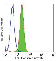
Human cervical cancer cell line HeLa was stained with EGFR (clone AY13) Alexa Fluor® 488 (filled histogram) or mouse IgG1, κ Alexa Fluor® 488 isotype control (open histogram). -

HeLa cells were fixed with 1% paraformaldehyde (PFA) and then stained with 10 µg/ml of anti-human EGFR (clone AY13) Alexa Fluor® 488 (green) for 3 hours at room temperature. Nuclei were counterstained with DAPI (blue). The image was captured by 40X objective.
| Cat # | Size | Price | Quantity Check Availability | Save | ||
|---|---|---|---|---|---|---|
| 352907 | 25 tests | £89 | ||||
| 352908 | 100 tests | £238 | ||||
Epidermal growth factor receptor (EGFR) is a transmembrane glycoprotein and member of the protein kinase superfamily that regulates cell growth and differentiation. EGFR binds EGF, TGF-α, amphiregulin, betacellulin, heparin-binding EGF-like growth factor, GP30, and vaccinia virus growth factor - all members of the EGF family. Ligand binding induces EGFR dimerization and autophosphorylation, initiating the MAPK, Akt, and JNK signaling pathways. EGFR is expressed by epithelial and endothelial cells and is frequently expressed by epithelial carcinomas.
Product DetailsProduct Details
- Verified Reactivity
- Human
- Antibody Type
- Monoclonal
- Host Species
- Mouse
- Immunogen
- Non-small cell lung cancer (NSCLC) cell line NCI-H322
- Formulation
- Phosphate-buffered solution, pH 7.2, containing 0.09% sodium azide and BSA (origin USA)
- Preparation
- The antibody was purified by affinity chromatography and conjugated with Alexa Fluor® 488 under optimal conditions.
- Concentration
- Lot-specific (to obtain lot-specific concentration and expiration, please enter the lot number in our Certificate of Analysis online tool.)
- Storage & Handling
- The antibody solution should be stored undiluted between 2°C and 8°C, and protected from prolonged exposure to light. Do not freeze.
- Application
-
FC - Quality tested
ICC - Verified - Recommended Usage
-
Each lot of this antibody is quality control tested by immunofluorescent staining with flow cytometric analysis. For flow cytometric staining, the suggested use of this reagent is 5 µL per million cells in 100 µL staining volume or 5 µL per 100 µL of whole blood.
* Alexa Fluor® 488 has a maximum emission of 519 nm when it is excited at 488 nm.
Alexa Fluor® and Pacific Blue™ are trademarks of Life Technologies Corporation.
View full statement regarding label licenses - Excitation Laser
-
Blue Laser (488 nm)
-
Application References
(PubMed link indicates BioLegend citation) -
- Yamaguchi M, et al. 2009. The 15th Annual Meeting Japan Society of Gene Therapy. p1056. Abstract 92.
- Product Citations
-
- RRID
-
AB_11124324 (BioLegend Cat. No. 352907)
AB_11126165 (BioLegend Cat. No. 352908)
Antigen Details
- Structure
- Member of the protein kinase superfamily; transmembrane glycoprotein; ligand binding induces dimerization and autophosphorylation
- Distribution
-
Epithelial cells and endothelial cells; frequently found in epithelial carcinomas
- Function
- Controls cell growth and differentiation
- Interaction
- MAPK, Akt, JNK
- Ligand/Receptor
- Members of the epidermal growth factor (EGF) family such as EGF, TGF-α, amphiregulin, betacellulin, heparin-binding EGF-like growth factor, GP30 and vaccinia virus growth factor
- Cell Type
- Endothelial cells, Epithelial cells
- Biology Area
- Cell Biology, Cell Cycle/DNA Replication, Immunology, Innate Immunity, Neuroscience, Synaptic Biology
- Molecular Family
- CD Molecules, Growth Factors
- Antigen References
-
1. da Cunha Santos G, et al. 2011. Annu. Rev. Pathol. 6:49.
2. Gusterson BA and Hunter KD. 2009. Lancet Oncol. 10:522.
3. Mano M and Humblet Y. 2008. Nat. Clin. Pract. Oncol. 5:415.
4. Pao W and Chmielecki J. 2010. Nat. Rev. Cancer 10:760. - Gene ID
- 1956 View all products for this Gene ID
- UniProt
- View information about EGFR on UniProt.org
Related Pages & Pathways
Pathways


Related FAQs
Other Formats
View All EGFR Reagents Request Custom Conjugation| Description | Clone | Applications |
|---|---|---|
| Purified anti-human EGFR | AY13 | FC,ICC |
| PE anti-human EGFR | AY13 | FC |
| APC anti-human EGFR | AY13 | FC |
| Alexa Fluor® 488 anti-human EGFR | AY13 | FC,ICC |
| Brilliant Violet 421™ anti-human EGFR | AY13 | FC,ICC |
| PE/Cyanine7 anti-human EGFR | AY13 | FC |
| PerCP/Cyanine5.5 anti-human EGFR | AY13 | FC |
| Alexa Fluor® 594 anti-human EGFR | AY13 | ICC,IHC-P |
| Alexa Fluor® 647 anti-human EGFR | AY13 | FC |
| Brilliant Violet 711™ anti-human EGFR | AY13 | FC |
| PE/Dazzle™ 594 anti-human EGFR | AY13 | FC |
| TotalSeq™-A0132 anti-human EGFR | AY13 | PG |
| Brilliant Violet 605™ anti-human EGFR | AY13 | FC |
| APC/Fire™ 750 anti-human EGFR | AY13 | FC |
| TotalSeq™-B0132 anti-human EGFR | AY13 | PG |
| TotalSeq™-C0132 anti-human EGFR | AY13 | PG |
| Biotin anti-human EGFR | AY13 | FC |
| TotalSeq™-D0132 anti-human EGFR | AY13 | PG |
| Brilliant Violet 650™ anti-human EGFR | AY13 | FC |
Customers Also Purchased

Compare Data Across All Formats
This data display is provided for general comparisons between formats.
Your actual data may vary due to variations in samples, target cells, instruments and their settings, staining conditions, and other factors.
If you need assistance with selecting the best format contact our expert technical support team.
-
Purified anti-human EGFR

HELA cells stained with purified EGFR (clone AY13, filled hi... 
HeLa cells were fixed with 2% PFA for ten minutes, permeabil... -
PE anti-human EGFR

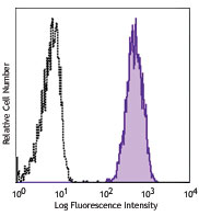
Human cervical cancer cell line HELA was stained with EGFR (... -
APC anti-human EGFR

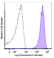
Human cervical cancer cell line HELA was stained with EGFR (... -
Alexa Fluor® 488 anti-human EGFR

Human cervical cancer cell line HeLa was stained with EGFR (... 
HeLa cells were fixed with 1% paraformaldehyde (PFA) and the... -
Brilliant Violet 421™ anti-human EGFR

Human cervical cancer cell line Hela was stained with EGFR (... 
HeLa cells were fixed with 1% paraformaldehyde solution and ... -
PE/Cyanine7 anti-human EGFR

Human cervical cancer cell line, Hela, was stained with EGFR... -
PerCP/Cyanine5.5 anti-human EGFR

HeLa cells were stained with EGFR (clone AY13) PerCP/Cyanine... -
Alexa Fluor® 594 anti-human EGFR

HeLa cells were fixed with 1% paraformaldehyde (PFA) and blo... 
Formalin-fixed paraffin-embedded human breast cancer tissue ... -
Alexa Fluor® 647 anti-human EGFR

Human cervical cancer cell line HELA was stained with EGFR (... -
Brilliant Violet 711™ anti-human EGFR

Human cervical cancer cell line HELA was stained with EGFR (... -
PE/Dazzle™ 594 anti-human EGFR

Human cervical cancer cell line HELA was stained with EGFR (... -
TotalSeq™-A0132 anti-human EGFR
-
Brilliant Violet 605™ anti-human EGFR

Human cervical cancer cell line HELA was stained with EGFR (... -
APC/Fire™ 750 anti-human EGFR

Human cervical cancer cell line HELA was stained with EGFR (... -
TotalSeq™-B0132 anti-human EGFR
-
TotalSeq™-C0132 anti-human EGFR
-
Biotin anti-human EGFR

Human cervical cancer cell line HELA was stained with biotin... -
TotalSeq™-D0132 anti-human EGFR
-
Brilliant Violet 650™ anti-human EGFR

Human cervical cancer cell line HeLa was stained with anti-h...


Follow Us