 Login / Register


- Clone
- 11B11 (See other available formats)
- Regulatory Status
- RUO
- Other Names
- Interleukin-4, Ia inducing factor (IaIF), B cell stimulating factor-1 (BSF-1), Hodgkin's cell growth factor (HCGF), Mast cell growth factor-2 (MCGF-2), Macrophage fusion factor (MFF), T cell growth factor-2 (TCGF-2)
- Isotype
- Rat IgG1, κ
- Ave. Rating
- Submit a Review
- Product Citations
- publications

-

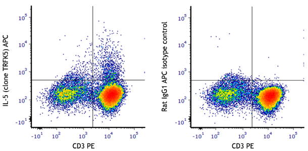
PMA+ionomycin-stimulated (in presence of Brefeldin A for 6 hours) Th2-polarized C57BL/6 mouse CD4+ T cells were surface stained with CD3 and then intracelluarly stained with IL-4 (clone 11B11) PE/Cyanine7 (left) or rat IgG1, κ PE/Cyanine7 isotype control (right).
| Cat # | Size | Price | Quantity Check Availability | Save | ||
|---|---|---|---|---|---|---|
| 504117 | 25 µg | 104€ | ||||
| 504118 | 100 µg | 268€ | ||||
IL-4 is a pleiotropic cytokine produced by activated T cells, mast cells, and basophils. IL-4 is a potent lymphoid cell growth factor which stimulates the growth and activation of certain B cells and T cells. IL-4 is important for regulation of T helper subset development.
Product DetailsProduct Details
- Verified Reactivity
- Mouse
- Antibody Type
- Monoclonal
- Host Species
- Rat
- Immunogen
- Partially purified native mouse IL-4
- Formulation
- Phosphate-buffered solution, pH 7.2, containing 0.09% sodium azide.
- Preparation
- The antibody was purified by affinity chromatography and conjugated with PE/Cyanine7 under optimal conditions.
- Concentration
- 0.2 mg/ml
- Storage & Handling
- The antibody solution should be stored undiluted between 2°C and 8°C, and protected from prolonged exposure to light. Do not freeze.
- Application
-
ICFC - Quality tested
- Recommended Usage
-
Each lot of this antibody is quality control tested by intracellular immunofluorescent staining with flow cytometric analysis. For flow cytometric staining, the suggested use of this reagent is =1.0 µg per million cells in 100 µl volume. It is recommended that the reagent be titrated for optimal performance for each application.
- Excitation Laser
-
Blue Laser (488 nm)
Green Laser (532 nm)/Yellow-Green Laser (561 nm)
- Application Notes
-
ELISA1,2,10,13 or ELISPOT5 Capture: The purified 11B11 antibody is useful as the capture antibody in a sandwich ELISA or ELISPOT assay, when used in conjunction with the biotinylated BVD6-24G2 antibody (Cat. No. 504202) as the detecting antibody and recombinant mouse IL-4 (Cat. No. 575609) as the standard. The LEAF™ purified antibody is suggested for ELISPOT capture.
Neutralization1-2,9,12: The 11B11 antibody can neutralize the bioactivity of natural or recombinant IL-4. The LEAF™ purified antibody (Endotoxin <0.1 EU/µg, Azide-Free, 0.2 µm filtered) is recommended for neutralization of mouse IL-4 bioactivity in vivo and in vitro (Cat. No. 504108).
Additional reported applications (for the relevant formats) include: immunoprecipitation16, immunohistochemical staining of formalin-fixed paraffin-embedded tissue sections8 and paraformaldehyde-fixed, saponin-treated frozen tissue sections6,7, and immunocytochemistry4.
Note: For testing mouse IL-4 in serum, plasma or supernatant, BioLegend's ELISA Max™ Sets (Cat. No. 431101 to 431106) are specially developed and recommended. - Additional Product Notes
-
BioLegend is in the process of converting the name PE/Cy7 to PE/Cyanine7. The dye molecule remains the same, so you should expect the same quality and performance from our PE/Cyanine7 products. Please contact Technical Service if you have any questions.
-
Application References
(PubMed link indicates BioLegend citation) -
- Shirai A, et al. 1994. Cytokine 6:329. (ELISA, Neut)
- Abrams J. 1995. Curr. Prot. Immunol. John Wiley and Sons New York. Unit 6.20. (ELISA, Neut)
- Assenmacher M, et al. 1994. Eur. J. Immunol. 24:1097.
- Openshaw P, et al. 1995. J. Exp. Med. 182:1357. (ICC)
- Klinman D, et al. 1994. Curr. Prot. Immunol. John Wiley and Sons New York. Unit 6.19. (ELISA Capture)
- Litton M, et al. 1994. J. Immunol. Methods 175:47. (IHC)
- Andersson U, et al. 1999. Detection and quantification of gene expression. New York:Springer-Verlag. (IHC)
- Fan WY, et al. 2001. Exp. Biol. Med. 226:1045. (IHC)
- Hara M, et al. 2001. J. Immunol. 166:3789. (Neut)
- Dzhagalov I, et al. 2007. J. Immunol. 178:2113. (ELISA)
- Lawson BR, et al. 2007. J. Immunol. 178:5366.
- Wang W, et al. 2007. J. Immunol. 178:4885. (Neut)
- Xu G, et al. 2007. J. Immunol. 179:5358. (ELISA) PubMed
- Ohnmacht C, et al. 2008. Blood 113:2816. PubMed
- Charles N, et al. 2010. Nat. Med. 16:701. (FC) PubMed
- Zavorotinskaya T, et al. 2003. Mol. Ther. 7:155. (IP)
- Product Citations
-
- RRID
-
AB_10895747 (BioLegend Cat. No. 504117)
AB_10898116 (BioLegend Cat. No. 504118)
Antigen Details
- Structure
- Cytokine; 15-19 kD (Mammalian)
- Bioactivity
- Differentiation of naïve CD4+ T cells to the TH2 type, proliferation/differentiation of activated B cells, expression of class II MHC antigens, and of low affinity IgE receptors in resting B cells
- Cell Sources
- Mast cells, T cells, bone marrow stromal cells
- Cell Targets
- B cells, T cells, monocytes, endothelial cells, fibroblasts
- Receptors
- Heterodimer IL-4Rα (CD124); γ-subunit (CD132) in common with IL-2R, IL-7R, IL-13R, IL-15R
- Cell Type
- Tregs
- Biology Area
- Immunology
- Molecular Family
- Cytokines/Chemokines
- Antigen References
-
1. Fitzgerald K, et al. Eds. 2001. The Cytokine FactsBook. Academic Press San Diego.
2. Boulay J, et al. 1992. Curr. Opin. Immunol. 4:294.
3. Dullens H, et al. 1991. In vivo 5:567.
4. Paul W. 1991. Blood 77:1859. - Regulation
- Upregulated by IL-2, platelet activating factor; downregulated by TGF-β
- Gene ID
- 16189 View all products for this Gene ID
- UniProt
- View information about IL-4 on UniProt.org
Related Pages & Pathways
Pathways


Related FAQs
Other Formats
View All IL-4 Reagents Request Custom Conjugation| Description | Clone | Applications |
|---|---|---|
| APC anti-mouse IL-4 | 11B11 | ICFC |
| PE anti-mouse IL-4 | 11B11 | ICFC |
| Purified anti-mouse IL-4 | 11B11 | ELISA Capture,CyTOF®,IP,IHC,ICC |
| Alexa Fluor® 488 anti-mouse IL-4 | 11B11 | ICFC |
| Alexa Fluor® 647 anti-mouse IL-4 | 11B11 | ICFC |
| PE/Cyanine7 anti-mouse IL-4 | 11B11 | ICFC |
| Brilliant Violet 421™ anti-mouse IL-4 | 11B11 | ICFC |
| Ultra-LEAF™ Purified anti-mouse IL-4 | 11B11 | ELISA Capture,CyTOF®,ELISPOT Capture,Neut,ICC,IP,IHC |
| PerCP/Cyanine5.5 anti-mouse IL-4 | 11B11 | ICFC |
| Brilliant Violet 605™ anti-mouse IL-4 | 11B11 | ICFC |
| Purified anti-mouse IL-4 (Maxpar® Ready) | 11B11 | ELISA Capture,CyTOF® |
| PE/Dazzle™ 594 anti-mouse IL-4 | 11B11 | ICFC |
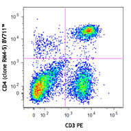
| Brilliant Violet 711™ anti-mouse IL-4 | 11B11 | ICFC |
| APC/Fire™ 750 anti-mouse IL-4 | 11B11 | ICFC |
Customers Also Purchased
Compare Data Across All Formats
This data display is provided for general comparisons between formats.
Your actual data may vary due to variations in samples, target cells, instruments and their settings, staining conditions, and other factors.
If you need assistance with selecting the best format contact our expert technical support team.
-
APC anti-mouse IL-4

PMA+ionomycin-stimulated (6 hours, in presence of brefeldin ... 
-
PE anti-mouse IL-4

PMA+ionomycin-stimulated (6 hours, in presence of brefeldin ... 
-
Purified anti-mouse IL-4

-
Alexa Fluor® 488 anti-mouse IL-4

PMA+ionomycin-stimulated (6 hours, in presence of brefeldin ... 
-
Alexa Fluor® 647 anti-mouse IL-4

PMA+ionomycin-stimulated (6 hours, in presence of brefeldin ... 
-
PE/Cyanine7 anti-mouse IL-4

PMA+ionomycin-stimulated (in presence of Brefeldin A for 6 h... -
Brilliant Violet 421™ anti-mouse IL-4

PMA+ionomycin-stimulated (6 hours, in presence of brefeldin ... 
-
Ultra-LEAF™ Purified anti-mouse IL-4
-
PerCP/Cyanine5.5 anti-mouse IL-4

Th2-polarized cells from C57BL/6 mouse T cells were stimulat... -
Brilliant Violet 605™ anti-mouse IL-4

PMA+ionomycin-stimulated (6 hours, in presence of brefeldin ... 
-
Purified anti-mouse IL-4 (Maxpar® Ready)

C57BL/6 mouse splenocytes were incubated for 20 hours in med... 
-
PE/Dazzle™ 594 anti-mouse IL-4

PMA+ionomycin-stimulated (6 hours, in the presence of brefel... 
-
Brilliant Violet 711™ anti-mouse IL-4

PMA + ionomycin-stimulated (six hours, in presence of brefel... 
-
APC/Fire™ 750 anti-mouse IL-4

PMA+ionomycin-stimulated (6 hours, in presence of brefeldin ...


Follow Us