 Login / Register


- Clone
- S16009A (See other available formats)
- Regulatory Status
- RUO
- Other Names
- Prf1, Pfp, Pfn, Pfn-1
- Isotype
- Rat IgG2a, κ
- Ave. Rating
- Submit a Review
- Product Citations
- publications

-

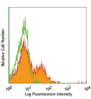
C57BL/6 splenocytes were stained with CD49b APC. Cells were fixed, permeabilized, and stained with PE anti-mouse perforin (clone S16009A, top) or PE rat IgG2a, κ isotype control (bottom). -

| Cat # | Size | Price | Quantity Check Availability | Save | ||
|---|---|---|---|---|---|---|
| 154305 | 25 µg | 113€ | ||||
| 154306 | 100 µg | 298€ | ||||
Perforin is a 70 kD cytolytic protein that is expressed in the cytoplasmic granules of cytotoxic T lymphocytes (CTLs) and natural killer (NK) cells. Perforin is one of the major effector molecules used by cytotoxic T cells and NK cells to mediate targeted cell lysis.
Product DetailsProduct Details
- Verified Reactivity
- Mouse
- Antibody Type
- Monoclonal
- Host Species
- Rat
- Immunogen
- Mouse perforin transfected cells.
- Formulation
- Phosphate-buffered solution, pH 7.2, containing 0.09% sodium azide.
- Preparation
- The antibody was purified by affinity chromatography and conjugated with PE under optimal conditions.
- Concentration
- 0.2 mg/ml
- Storage & Handling
- The antibody solution should be stored undiluted between 2°C and 8°C, and protected from prolonged exposure to light. Do not freeze.
- Application
-
ICFC - Quality tested
- Recommended Usage
-
Each lot of this antibody is quality control tested by intracellular immunofluorescent staining with flow cytometric analysis. For flow cytometric staining, the suggested use of this reagent is ≤ 0.5 µg per million cells in 100 µl volume. It is recommended that the reagent be titrated for optimal performance for each application.
- Excitation Laser
-
Blue Laser (488 nm)
Green Laser (532 nm)/Yellow-Green Laser (561 nm)
- Application Notes
-
Clone S16009A reacts with perforin from both C57BL/6 and BALB/c strains.
- Product Citations
-
- RRID
-
AB_2721638 (BioLegend Cat. No. 154305)
AB_2721639 (BioLegend Cat. No. 154306)
Antigen Details
- Structure
- 70 kD Protein that oligomerizes for pore formation.
- Distribution
-
CTL, NK (Cytoplasmic Granules)
- Function
- Target cell lysis
- Cell Type
- NK cells, T cells
- Biology Area
- Immunology
- Molecular Family
- Cytokines/Chemokines
- Antigen References
-
- Podack ER, et al. 1988. Immunol. Rev. 103:203.
- Konjar S, et al. 2010. Immunology. 131:257.
- Trapani JA, et al. 2002. Nat. Rev. Immunol. 2:735.
- Osinska I, et al. 2014. Cent. Eur. J. Immnol. 39:109.
- Gene ID
- 18646 View all products for this Gene ID
- UniProt
- View information about Perforin on UniProt.org
Related Pages & Pathways
Pathways


Related FAQs
- What type of PE do you use in your conjugates?
- We use R-PE in our conjugates.
Other Formats
View All Perforin Reagents Request Custom Conjugation| Description | Clone | Applications |
|---|---|---|
| APC anti-mouse Perforin | S16009A | ICFC |
| PE anti-mouse Perforin | S16009A | ICFC |
| Purified anti-mouse Perforin | S16009A | ICFC |
| FITC anti-mouse Perforin | S16009A | ICFC |
| PE/Dazzle™ 594 anti-mouse Perforin | S16009A | ICFC |
| APC/Fire™ 750 anti-mouse Perforin | S16009A | ICFC |
| Pacific Blue™ anti-mouse Perforin | S16009A | ICFC |
| Alexa Fluor® 647 anti-mouse Perforin | S16009A | ICFC |
| Brilliant Violet 421™ anti-mouse Perforin | S16009A | ICFC |
Customers Also Purchased
Compare Data Across All Formats
This data display is provided for general comparisons between formats.
Your actual data may vary due to variations in samples, target cells, instruments and their settings, staining conditions, and other factors.
If you need assistance with selecting the best format contact our expert technical support team.
-
APC anti-mouse Perforin

C57BL/6 splenocytes were stained with CD49b PE. Cells were ... 
-
PE anti-mouse Perforin

C57BL/6 splenocytes were stained with CD49b APC. Cells were... 
-
Purified anti-mouse Perforin

C57BL/6 splenocytes were fixed, permeabilized, and stained w... -
FITC anti-mouse Perforin

C57 splenocytes were stained with anti-mouse CD49b APC, fixe... -
PE/Dazzle™ 594 anti-mouse Perforin

C57BL/6 splenocytes were stained with anti-mouse CD49b (clon... -
APC/Fire™ 750 anti-mouse Perforin

C57BL/6 splenocytes were stained with anti-mouse CD49b APC. ... -
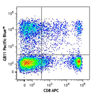
Pacific Blue™ anti-mouse Perforin

C57 splenocytes were stained with anti-mouse CD49b FITC, fix... -
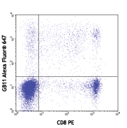
Alexa Fluor® 647 anti-mouse Perforin

C57 splenocytes were stained with anti-mouse CD49b PE, fixed... -
Brilliant Violet 421™ anti-mouse Perforin

C57BL/6 splenocytes were stained with anti-mouse CD49b APC. ...


Follow Us