 Login / Register


- Clone
- 3A6 (See other available formats)
- Regulatory Status
- RUO
- Workshop
- HCDM listed
- Other Names
- CD6 ligand, Activated Leukocyte Cell Adhesion Molecule (ALCAM)
- Isotype
- Mouse IgG1, κ
- Ave. Rating
- Submit a Review
- Product Citations
- publications

-

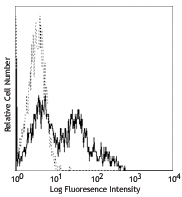
PHA-stimulated human peripheral blood lymphocytes (3 days) stained with 3A6 PE -

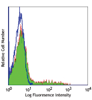
Human peripheral blood monocytes stained with 3A6 PE
| Cat # | Size | Price | Quantity Check Availability | Save | ||
|---|---|---|---|---|---|---|
| 343903 | 25 tests | 112€ | ||||
| 343904 | 100 tests | 225€ | ||||
CD166, also known as the CD6 ligand or the Activated Leukocyte Cell Adhesion Molecule (ALCAM), is a 100-105 kD transmembrane glycoprotein. It belongs to the Ig superfamily of proteins and expressed on activated T cells, activated monocytes, epithelial cells, fibroblasts, and neurons. CD166 plays an important role in mediating adhesion interactions between thymic epithelial cells and CD6+ cells during intrathymic T cell development. Recently CD166 has also been used as a potential cancer stem cell marker. The antibody reacts with human activated leukocyte cell adhesion molecule (ALCAM).
Product DetailsProduct Details
- Verified Reactivity
- Human
- Reported Reactivity
- African Green, Baboon, Cynomolgus, Rhesus
- Antibody Type
- Monoclonal
- Host Species
- Mouse
- Immunogen
- Cultured human thymic epithelial cells
- Formulation
- Phosphate-buffered solution, pH 7.2, containing 0.09% sodium azide and BSA (origin USA)
- Preparation
- The antibody was purified by affinity chromatography, and conjugated with PE under optimal conditions.
- Concentration
- Lot-specific (to obtain lot-specific concentration and expiration, please enter the lot number in our Certificate of Analysis online tool.)
- Storage & Handling
- The antibody solution should be stored undiluted between 2°C and 8°C, and protected from prolonged exposure to light. Do not freeze.
- Application
-
FC - Quality tested
- Recommended Usage
-
Each lot of this antibody is quality control tested by immunofluorescent staining with flow cytometric analysis. For flow cytometric staining, the suggested use of this reagent is 5 µl per million cells in 100 µl staining volume or 5 µl per 100 µl of whole blood.
- Excitation Laser
-
Blue Laser (488 nm)
Green Laser (532 nm)/Yellow-Green Laser (561 nm)
- Application Notes
-
Additional reported applications (for the relevant formats) include: immunohistochemical staining of paraffin-embedded tissue sections and immunofluorescence.1
-
Application References
(PubMed link indicates BioLegend citation) -
- Pretzel D, et al. 2011. Arthritis Res. Ther. 13:R64. (IHC, IF, FC)
- Product Citations
-
- RRID
-
AB_2289303 (BioLegend Cat. No. 343903)
AB_2289302 (BioLegend Cat. No. 343904)
Antigen Details
- Structure
- A type I transmembrane glycoprotein belonging to the Ig superfamily of proteins.
- Distribution
-
CD166 is expressed on activated T cells, activated monocytes, epithelial cells, fibroblasts, and neurons.
- Function
- Play an important role in mediating adhesion interactions between thymic epithelial cells and CD6+ cells during intrathymic T cell development.
- Ligand/Receptor
- CD6
- Cell Type
- Epithelial cells, Fibroblasts, Mesenchymal Stem Cells, Monocytes, Neurons, T cells
- Biology Area
- Immunology, Stem Cells
- Molecular Family
- Adhesion Molecules, CD Molecules
- Antigen References
-
1. Aruffo A, et al. 1997. Immunol Today. 18(10):498
2. Patel DD, et al. 1995. J. Exp. Med. 181:2213
3. Bowen MA, et al. 1995. J. Exp. Med. 181:1563
4. Horst D, et al. 2009. Cancer Invest. 22:1 - Gene ID
- 214 View all products for this Gene ID
- UniProt
- View information about CD166 on UniProt.org
Related Pages & Pathways
Pathways


Related FAQs
- What type of PE do you use in your conjugates?
- We use R-PE in our conjugates.
Other Formats
View All CD166 Reagents Request Custom Conjugation| Description | Clone | Applications |
|---|---|---|
| Purified anti-human CD166 | 3A6 | FC,IHC-P,ICC |
| PE anti-human CD166 | 3A6 | FC |
| APC/Fire™ 750 anti-human CD166 | 3A6 | FC |
| PE/Cyanine7 anti-human CD166 | 3A6 | FC |
| PerCP/Cyanine5.5 anti-human CD166 | 3A6 | FC |
| APC anti-human CD166 | 3A6 | FC |
| TotalSeq™-A1015 anti-human CD166 | 3A6 | PG |
| TotalSeq™-D1015 anti-human CD166 Antibody | 3A6 | PG |
| TotalSeq™-C1015 anti-human CD166 | 3A6 | PG |
| TotalSeq™-B1015 anti-human CD166 | 3A6 | PG |
| Brilliant Violet 421™ anti-human CD166 | 3A6 | FC |
| KIRAVIA Blue 520™ anti-human CD166 | 3A6 | FC |
| PE/Dazzle™ 594 anti-human CD166 | 3A6 | FC |
| Brilliant Violet 785™ anti-human CD166 | 3A6 | FC |
| Alexa Fluor® 647 anti-human CD166 | 3A6 | FC |
Customers Also Purchased
Compare Data Across All Formats
This data display is provided for general comparisons between formats.
Your actual data may vary due to variations in samples, target cells, instruments and their settings, staining conditions, and other factors.
If you need assistance with selecting the best format contact our expert technical support team.
-
Purified anti-human CD166

PHA-stimulated human peripheral blood lymphocytes (3 days) s... 
Human peripheral blood monocytes stained with purified 3A6 c... -
PE anti-human CD166
PHA-stimulated human peripheral blood lymphocytes (3 days) s...
Human peripheral blood monocytes stained with 3A6 PE -
APC/Fire™ 750 anti-human CD166

PHA-stimulated (3 day) human peripheral blood lymphocytes we... -
PE/Cyanine7 anti-human CD166

PHA-stimulated (3 day) human peripheral blood lymphocytes we... -
PerCP/Cyanine5.5 anti-human CD166

PHA-stimulated (3 day) human peripheral blood lymphocytes we... -
APC anti-human CD166

PHA-stimulated (3 day) human peripheral blood lymphocytes we... -
TotalSeq™-A1015 anti-human CD166
-
TotalSeq™-D1015 anti-human CD166 Antibody
-
TotalSeq™-C1015 anti-human CD166
-
TotalSeq™-B1015 anti-human CD166
-
Brilliant Violet 421™ anti-human CD166

PHA-stimulated (3 day) human peripheral blood lymphocytes we... -
KIRAVIA Blue 520™ anti-human CD166

PHA-stimulated (3 day) human peripheral blood lymphocytes we... -
PE/Dazzle™ 594 anti-human CD166

PHA-stimulated (3 day) human peripheral blood lymphocytes we... -
Brilliant Violet 785™ anti-human CD166

PHA-stimulated (3 day) human peripheral blood lymphocytes we... -
Alexa Fluor® 647 anti-human CD166

PHA-stimulated (3 day) human peripheral blood lymphocytes we...


Follow Us