 Login / Register


- Clone
- X54-5/7.1 (See other available formats)
- Regulatory Status
- RUO
- Other Names
- FcRI
- Isotype
- Mouse IgG1, κ
- Ave. Rating
- Submit a Review
- Product Citations
- publications

-

C57BL/6 mouse bone marrow cells were stained with CD11b PE and CD64 (clone X54-5/7.1) APC (top) or mouse IgG1, κ APC isotype control (bottom). Data shown was gated on the myeloid population. -

| Cat # | Size | Price | Quantity Check Availability | Save | ||
|---|---|---|---|---|---|---|
| 139305 | 25 µg | 86€ | ||||
| 139306 | 100 µg | 214€ | ||||
CD64 is a 72 kD single chain type I glycoprotein also known as FcγRI and FcRI. CD64 is a member of the immunoglobulin superfamily and is expressed on monocytes/macrophages, dendritic cells, and mast cells. The expression can be upregulated by IFN-γ stimulation. CD64 binds IgG immune complex. It plays a role in antigen capture, phagocytosis of IgG/antigen complexes, and antibody-dependent cellular cytotoxicity (ADCC).
Product DetailsProduct Details
- Verified Reactivity
- Mouse
- Antibody Type
- Monoclonal
- Host Species
- Mouse
- Immunogen
- BALB/c mouse FcγRI-human IgG Fc fusion protein.
- Formulation
- Phosphate-buffered solution, pH 7.2, containing 0.09% sodium azide.
- Preparation
- The antibody was purified by affinity chromatography and conjugated with APC under optimal conditions.
- Concentration
- 0.2 mg/ml
- Storage & Handling
- The antibody solution should be stored undiluted between 2°C and 8°C, and protected from prolonged exposure to light. Do not freeze.
- Application
-
FC - Quality tested
- Recommended Usage
-
Each lot of this antibody is quality control tested by immunofluorescent staining with flow cytometric analysis. For flow cytometric staining, the suggested use of this reagent is ≤1.0 µg per million cells in 100 µl volume. It is recommended that the reagent be titrated for optimal performance for each application.
- Excitation Laser
-
Red Laser (633 nm)
- Application Notes
-
The X54-5/7.1 antibody reacts with mouse strains carrying CD64a and b alleles but not CD64d. X54-5/7.1 recognizes a conformational determinant formed between domains 2 and 3. Additional reported application (for relevant formats) include: immunoprecipitation1, and spatial biology (IBEX)5,6. Clone X54-5/7.1 is not found to be useful for Western blots1.
-
Application References
(PubMed link indicates BioLegend citation) -
- Tan PS, et al. 2003. J. Immunol. 170:2549. (IP)
- Ingersoll MA, et al. 2010. Blood 115:e10. (FC)
- Ozeri E, et al. 2012. J. Immunol. 189:146. PubMed
- Richardson ML, et al. 2014. PLoS Negl Trop Dis. 8:2825. PubMed
- Radtke AJ, et al. 2020. Proc Natl Acad Sci U S A. 117:33455-65. (SB) PubMed
- Radtke AJ, et al. 2022. Nat Protoc. 17:378-401. (SB) PubMed
- Product Citations
-
- RRID
-
AB_11219205 (BioLegend Cat. No. 139305)
AB_11219391 (BioLegend Cat. No. 139306)
Antigen Details
- Structure
- Ig superfamily, type I glycoprotein, 72 kD
- Distribution
-
Monocytes, macrophages, mast cells, dendritic cells
- Function
- Phagocytosis, ADCC
- Ligand/Receptor
- IgG
- Cell Type
- Dendritic cells, Macrophages, Mast cells, Monocytes
- Biology Area
- Immunology, Innate Immunity
- Molecular Family
- CD Molecules, Fc Receptors
- Gene ID
- 14129 View all products for this Gene ID
- UniProt
- View information about CD64 on UniProt.org


Related FAQs
Other Formats
View All CD64 Reagents Request Custom Conjugation| Description | Clone | Applications |
|---|---|---|
| Purified anti-mouse CD64 (FcγRI) | X54-5/7.1 | FC,IP |
| PE anti-mouse CD64 (FcγRI) | X54-5/7.1 | FC |
| APC anti-mouse CD64 (FcγRI) | X54-5/7.1 | FC |
| PerCP/Cyanine5.5 anti-mouse CD64 (FcγRI) | X54-5/7.1 | FC |
| Brilliant Violet 421™ anti-mouse CD64 (FcγRI) | X54-5/7.1 | FC |
| Brilliant Violet 711™ anti-mouse CD64 (FcγRI) | X54-5/7.1 | FC |
| PE/Cyanine7 anti-mouse CD64 (FcγRI) | X54-5/7.1 | FC |
| FITC anti-mouse CD64 (FcγRI) | X54-5/7.1 | FC |
| Biotin anti-mouse CD64 (FcγRI) | X54-5/7.1 | FC |
| PE/Dazzle™ 594 anti-mouse CD64 (FcγRI) | X54-5/7.1 | FC |
| Alexa Fluor® 647 anti-mouse CD64 (FcγRI) | X54-5/7.1 | FC,SB |
| Brilliant Violet 605™ anti-mouse CD64 (FcγRI) | X54-5/7.1 | FC |
| TotalSeq™-A0202 anti-mouse CD64 (FcγRI) | X54-5/7.1 | PG |
| TotalSeq™-C0202 anti-mouse CD64 (FcγRI) | X54-5/7.1 | PG |
| TotalSeq™-B0202 anti-mouse CD64 (FcγRI) | X54-5/7.1 | PG |
| PE/Cyanine5 anti-mouse CD64 (FcγRI) | X54-5/7.1 | FC |
| APC/Fire™ 750 anti-mouse CD64 (FcγRI) | X54-5/7.1 | FC |
| Brilliant Violet 510™ anti-mouse CD64 (FcγRI) | X54-5/7.1 | FC |
Customers Also Purchased
Compare Data Across All Formats
This data display is provided for general comparisons between formats.
Your actual data may vary due to variations in samples, target cells, instruments and their settings, staining conditions, and other factors.
If you need assistance with selecting the best format contact our expert technical support team.
-
Purified anti-mouse CD64 (FcγRI)

C57BL/6 bone marrow cells stained with CD11b FITC and purifi... 
C57BL/6 bone marrow cells stained with CD11b FITC and mouse ... -
PE anti-mouse CD64 (FcγRI)
C57BL/6 bone marrow cells stained with CD11b FITC and X54-5/...
C57BL/6 bone marrow cells stained with CD11b FITC and mouse ... -
APC anti-mouse CD64 (FcγRI)

C57BL/6 mouse bone marrow cells were stained with CD11b PE a... 
-
PerCP/Cyanine5.5 anti-mouse CD64 (FcγRI)

C57BL/6 mouse bone marrow cells were stained with CD11b APC ... -
Brilliant Violet 421™ anti-mouse CD64 (FcγRI)

C57BL/6 mouse bone marrow cells were stained with CD11b APC ... 
-
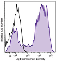
Brilliant Violet 711™ anti-mouse CD64 (FcγRI)

C57BL/6 mouse bone marrow cells were stained with CD11b FITC... 
-
PE/Cyanine7 anti-mouse CD64 (FcγRI)

C57BL/6 mouse bone marrow cells were stained with CD11b APC ... -
FITC anti-mouse CD64 (FcγRI)

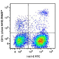
C57BL/6 mouse bone marrow cells were stained with CD11b (clo... 
-
Biotin anti-mouse CD64 (FcγRI)

C57BL/6 mouse bone marrow cells were stained with CD11b (clo... 
-
PE/Dazzle™ 594 anti-mouse CD64 (FcγRI)

C57BL/6 mouse bone marrow cells were stained with CD11b (clo... 
-
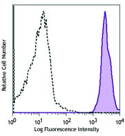
Alexa Fluor® 647 anti-mouse CD64 (FcγRI)

C57BL/6 mouse bone marrow cells were stained with CD11b (clo... 

Mice were injected subcutaneously with sheep red blood cells... -
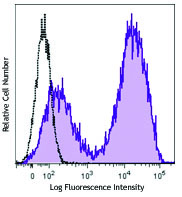
Brilliant Violet 605™ anti-mouse CD64 (FcγRI)

C57BL/6 mouse bone marrow cells were stained with CD11b APC ... 
-
TotalSeq™-A0202 anti-mouse CD64 (FcγRI)
-
TotalSeq™-C0202 anti-mouse CD64 (FcγRI)
-
TotalSeq™-B0202 anti-mouse CD64 (FcγRI)
-
PE/Cyanine5 anti-mouse CD64 (FcγRI)

C57BL/6 mouse bone marrow cells were stained with anti-mouse... -
APC/Fire™ 750 anti-mouse CD64 (FcγRI)

C57BL/6 mouse bone marrow cells were stained with anti-mouse... -
Brilliant Violet 510™ anti-mouse CD64 (FcγRI)

C57BL/6 mouse bone marrow was stained with anti-mouse/human ...


Follow Us