 Login / Register


- Clone
- TDP2H4 (See other available formats)
- Regulatory Status
- RUO
- Other Names
- TAR DNA-binding protein-43, TDP 43, TARDBP, ALS10
- Previously
-
Covance Catalog# SIG-39854
- Isotype
- Rat IgG2a
- Ave. Rating
- Submit a Review
- Product Citations
- publications

-

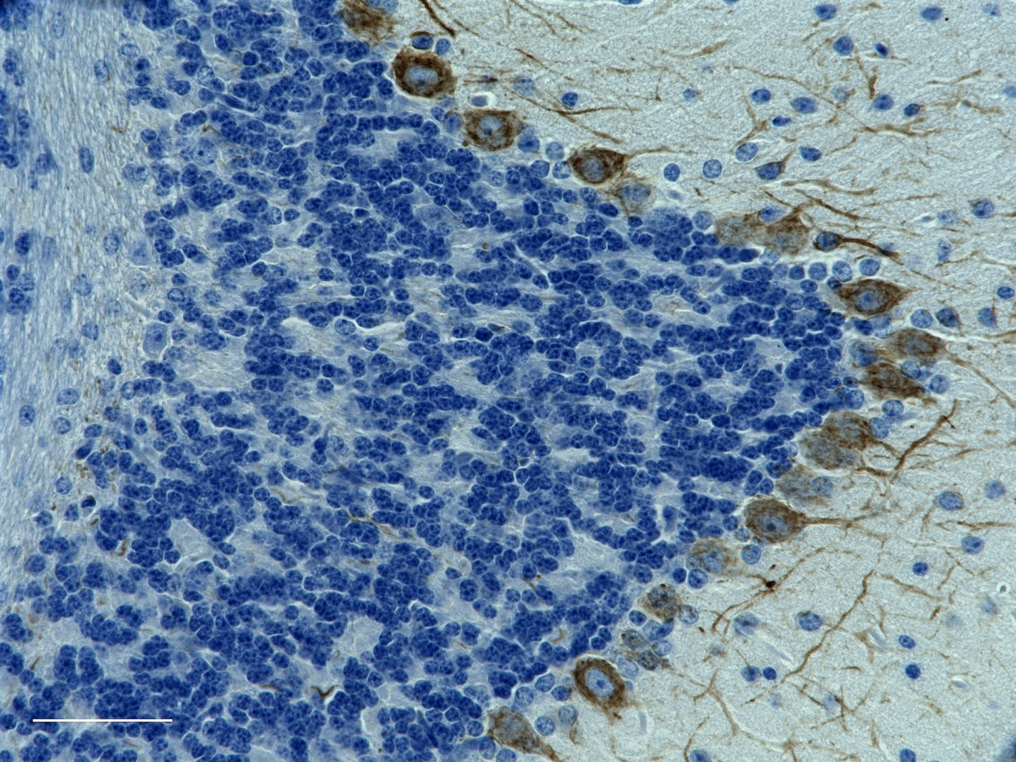
IHC staining of anti-TDP43 antibody (clone TDP2H4) on formalin-fixed paraffin-embedded rat (A) and mouse (B) brain tissues. Following antigen retrieval using Sodium Citrate H.I.E.R and 0.25% Triton X-100, tissues were incubated with 2 µg/ml of the primary antibody overnight at 4°C. After incubation with biotinylated anti-rat antibody, the tissue was labeled with BioLegend’s HRP labeling reagent followed by addition of DAB chromogen (Cat. No. 926506) for detection and hematoxylin counterstaining, according to the protocol provided. The images were captured with a 40X objective. Scale bar 50 µm. -

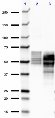
Western blot of anti-TDP43 antibody (clone TDP2H4). Lane 1: Molecular weight marker; Lane 2: 20 µg of human brain lysate; Lane 3: 20 µg of mouse brain lysates; Lane 4: 20 µg of rat brain lysates; Lane 5: 20 µg of Hela lysates. The blot was incubated with 1 µg/ml of the primary antibody overnight at 4°C, followed by the incubation with HRP-labeled goat anti-rat IgG (Cat. No. 405405). -

Western blot of anti-TDP43 antibody (clone TDP2H4). Lane 1: Molecular weight marker; Lane 2: 20 µg of human cerebellum lysate; Lane 3: 20 µg of human brain nuclear fraction. The blot was incubated with 1 µg/ml of the primary antibody overnight at 4°C, followed by the incubation with HRP-labeled goat anti-rat IgG (Cat. No. 405405). The anti-α-tubulin antibody (Cat. No. 909601) was used as a loading control. -

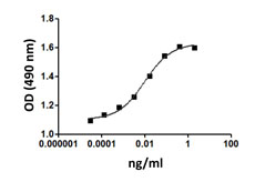
Direct ELISA of purified anti-TDP43 antibody (clone TDP2H4) binding to plate-immobilized full-length, N-terminal fragment (a.a. 1-207), or C-terminal fragment (a.a 208-414) of TDP43. ELISA was performed by coating wells with 100 ng of each protein. The wells were then incubated with the primary antibody at 37°C for 45 minutes, followed by incubation with horseradish peroxidase labeled goat anti-rat secondary antibody. TMB (3, 3', 5, 5' tetramethylbenzidine) was used as the detection system.
| Cat # | Size | Price | Quantity Check Availability | Save | ||
|---|---|---|---|---|---|---|
| 808301 | 100 µL | 296€ | ||||
TDP43, also known as TARDBP, was originally identified as a protein that binds to the transactivation response (TAR) sequence found in the long terminal repeat of the HIV-1 virus genome. TDP43 contains two copies of a ~90 amino acid RNA recognition motif (RRM) found in many proteins, which bind single stranded RNA. Recent research has shown that hyperphosphorylated, ubiquitinated, and N-terminally truncated TDP43 is the pathological hallmark lesion in most familial and sporadic forms of FTLD-U and ALS (1).
Product DetailsProduct Details
- Verified Reactivity
- Human, Mouse, Rat
- Antibody Type
- Monoclonal
- Host Species
- Rat
- Immunogen
- This antibody was raised against a nonphosphorylated peptide corresponding to the C-terminal domain of human TDP43, which includes serine residues 409 and 410.
- Formulation
- Phosphate-buffered solution.
- Preparation
- The antibody was purified by affinity chromatography.
- Concentration
- 1.0 mg/ml
- Storage & Handling
- The antibody solution should be stored undiluted between 2°C and 8°C. Please note the storage condition for this antibody has been changed from -20°C to between 2°C and 8°C. You can also check your vial or your CoA to find the most accurate storage condition for this antibody.
- Application
-
IHC-P - Quality tested
WB, Direct ELISA - Verified - Recommended Usage
-
Each lot of this antibody is quality control tested by formalin-fixed paraffin-embedded immunohistochemical staining. For immunohistochemistry, a dilution of 1:500 is suggested. For Western blotting, the suggested use of this reagent is 1.0 – 5.0 µg per ml. For Direct ELISA application, a concentration range of 10 ng - 1.0 µg/mL is recommended. It is recommended that the reagent be titrated for optimal performance for each application.
- Application Notes
-
This antibody is effective in immunoblotting (WB), immunohistochemistry (IHC), and Direct ELISA.
Expected Staining Pattern: Nuclear -
Application References
(PubMed link indicates BioLegend citation) -
- Neumann M. 2009. Int. J. Mol. Sci. 10.3390/ijms10010232.
- RRID
-
AB_2564740 (BioLegend Cat. No. 808301)
Antigen Details
- Structure
- TDP43 is a 414 amino acid protein with an apparent molecular mass of 45 kD.
- Biology Area
- Cell Biology, Neurodegeneration, Neuroscience, Protein Misfolding and Aggregation
- Molecular Family
- TDP43
- Gene ID
- 23435 View all products for this Gene ID 230908 View all products for this Gene ID 298648 View all products for this Gene ID
- UniProt
- View information about TDP43 on UniProt.org


Related FAQs
Other Formats
View All TDP43 Reagents Request Custom Conjugation| Description | Clone | Applications |
|---|---|---|
| Purified anti-TDP43 | TDP2H4 | IHC-P,WB,Direct ELISA |
| Biotin anti-TDP43 | TDP2H4 | WB,IHC-P |
Customers Also Purchased
Compare Data Across All Formats
This data display is provided for general comparisons between formats.
Your actual data may vary due to variations in samples, target cells, instruments and their settings, staining conditions, and other factors.
If you need assistance with selecting the best format contact our expert technical support team.
-
Purified anti-TDP43

IHC staining of anti-TDP43 antibody (clone TDP2H4) on formal... 
Western blot of anti-TDP43 antibody (clone TDP2H4). Lane 1: ... 
Western blot of anti-TDP43 antibody (clone TDP2H4). Lane 1: ... 
Direct ELISA of purified anti-TDP43 antibody (clone TDP2H4) ... -
Biotin anti-TDP43

Western blot of Biotin anti-TDP43 antibody (clone TDP2H4). L... 
IHC staining of Biotin anti-TDP43 antibody (clone TDP2H4) on... 
IHC staining of Biotin anti-TDP43 antibody (clone TDP2H4) on...


Follow Us