 Login / Register


- Clone
- SHM16 (See other available formats)
- Regulatory Status
- RUO
- Other Names
- Ephrin type-A receptor 2 (EPHA2), Epithelial cell kinase, Tyrosine-protein kinase receptor ECK, Myk2, Sek2, EPH receptor A2, eEpithelial cell receptor protein tyrosine kinase, Protein tyrosine kinase, Soluble EPHA2 variant 1
- Isotype
- Mouse IgG2b, κ
- Ave. Rating
- Submit a Review
- Product Citations
- publications

-

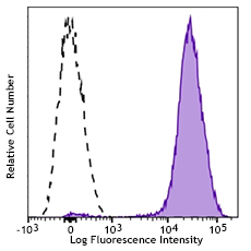
Human melanoma cell line, A375, was stained with EphA2 (clone SHM16) PE (filled histogram) or mouse IgG2b, κ PE isotype control (open histogram).
| Cat # | Size | Price | Quantity Check Availability | Save | ||
|---|---|---|---|---|---|---|
| 356803 | 25 tests | 117€ | ||||
| 356804 | 100 tests | 268€ | ||||
Ephrin receptor A2 (EphA2), also known as Eck, is a 130 kD type I transmembrane glycoprotein which belongs to the ephrin receptor subfamily of the protein-tyrosine kinase family. EphA2 is expressed on epithelial cells, keratinocytes, dendritic/Langerhans cells and melanocytes. It has also been observed in breast, cervical, ovarian, prostate, colon, skin and lung cancers. EphA2 is an oncoprotein which is an essential survival factor for many melanomas. EphA2 plays an important role in angiogenesis and tumor neovascularization through interaction with ephrin-A1, -A3, -A4 and -A5.
Product DetailsProduct Details
- Verified Reactivity
- Human
- Antibody Type
- Monoclonal
- Host Species
- Mouse
- Formulation
- Phosphate-buffered solution, pH 7.2, containing 0.09% sodium azide and BSA (origin USA)
- Preparation
- The antibody was purified by affinity chromatography and conjugated with PE under optimal conditions.
- Concentration
- Lot-specific (to obtain lot-specific concentration and expiration, please enter the lot number in our Certificate of Analysis online tool.)
- Storage & Handling
- The antibody solution should be stored undiluted between 2°C and 8°C, and protected from prolonged exposure to light. Do not freeze.
- Application
-
FC - Quality tested
- Recommended Usage
-
Each lot of this antibody is quality control tested by immunofluorescent staining with flow cytometric analysis. For flow cytometric staining, the suggested use of this reagent is 5 µl per million cells in 100 µl staining volume or 5 µl per 100 µl of whole blood.
- Excitation Laser
-
Blue Laser (488 nm)
Green Laser (532 nm)/Yellow-Green Laser (561 nm)
- Product Citations
-
- RRID
-
AB_2561806 (BioLegend Cat. No. 356803)
AB_2561807 (BioLegend Cat. No. 356804)
Antigen Details
- Structure
- Type I transmembrane glycoprotein, 130 kD, protein- tyrosine kinase family
- Distribution
- Epithelium (keratinocytes, renal collecting duct cells, carcinomas), dendritic/Langerhans cells, melanocytes
- Function
- Angiogenesis and tumor neovascularization
- Ligand/Receptor
- Ephrin-A1, -A3, -A4, -A5
- Cell Type
- Epithelial cells, Langerhans cells, Dendritic cells
- Biology Area
- Angiogenesis, Apoptosis/Tumor Suppressors/Cell Death, Cell Adhesion, Cell Biology, Immunology, Neuroscience, Synaptic Biology
- Molecular Family
- Adhesion Molecules, Protein Kinases/Phosphatase
- Antigen References
-
1. Pandey A, et al. 1994. J. Biol. Chem. 269:30154.
2. Sulman E, et al. 1997. Genomics 40:371.
3. Pratt R, et al. 2002. Oncogene 21:7690.
4. Udayakumar D, et al. 2011. Oncogene 30:4921.
5. Brantley-Sieders D, et al. 2008. J. Clin. Invest. 118:64. - Gene ID
- 1969 View all products for this Gene ID
- UniProt
- View information about EphA2 on UniProt.org


Related FAQs
- What type of PE do you use in your conjugates?
- We use R-PE in our conjugates.
Other Formats
View All EphA2 Reagents Request Custom Conjugation| Description | Clone | Applications |
|---|---|---|
| Purified anti-human EphA2 | SHM16 | FC |
| PE anti-human EphA2 | SHM16 | FC |
Customers Also Purchased
Compare Data Across All Formats
This data display is provided for general comparisons between formats.
Your actual data may vary due to variations in samples, target cells, instruments and their settings, staining conditions, and other factors.
If you need assistance with selecting the best format contact our expert technical support team.
-
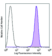
Purified anti-human EphA2

Human melanoma cell line, A375, was stained with purified Ep... -
PE anti-human EphA2

Human melanoma cell line, A375, was stained with EphA2 (clon...


Follow Us