 Login / Register


- Clone
- 2B8 (See other available formats)
- Regulatory Status
- RUO
- Other Names
- c-KIT, Stem Cell Factor Receptor (SCFR)
- Isotype
- Rat IgG2b, κ
- Ave. Rating
- Submit a Review
- Product Citations
- publications

-

-

-

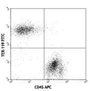
Confocal image of C57BL/6 mouse small intestine sample acquired using the IBEX method of highly multiplexed antibody-based imaging: EpCAM (cyan) in Cycle 1, B220 (purple) in Cycle 2, and CD117 (yellow) in Cycle 2. Tissues were prepared using ~1% (vol/vol) formaldehyde and a detergent. Following fixation, samples are immersed in 30% (wt/vol) sucrose for cryoprotection. Images are courtesy of Drs. Andrea J. Radtke and Ronald N. Germain of the Center for Advanced Tissue Imaging (CAT-I) in the National Institute of Allergy and Infectious Diseases (NIAID, NIH). -

Confocal image of human lymph node sample acquired using the IBEX method of highly multiplexed antibody-based imaging: CD35 (blue) in Cycle 6, CD117 (yellow) in Cycle 6, and CD66b (magenta) in Cycle 11. Tissues were prepared using ~1% (vol/vol) formaldehyde and a detergent. Following fixation, samples are immersed in 30% (wt/vol) sucrose for cryoprotection. Images are courtesy of Drs. Andrea J. Radtke and Ronald N. Germain of the Center for Advanced Tissue Imaging (CAT-I) in the National Institute of Allergy and Infectious Diseases (NIAID, NIH).
| Cat # | Size | Price | Quantity Check Availability | Save | ||
|---|---|---|---|---|---|---|
| 105827 | 125 µL | 132€ | ||||
| 105828 | 500 µL | 308€ | ||||
CD117 is a 145 kD immunoglobulin superfamily member also known as c-Kit and stem cell factor receptor (SCFR). It is a transmembrane tyrosine-kinase receptor that binds the c-Kit ligand (also known as steel factor, stem cell factor, and mast cell growth factor). CD117 is expressed on hematopoietic stem cells (including multipotent hematopoietic stem cells, progenitors committed to myeloid and/or erythroid lineages, and T and B cell precursors), mast cells, and acute myeloid leukemia (AML) cells. CD117 interaction with its ligand is critical for the development of hematopoietic stem cells.
Product DetailsProduct Details
- Verified Reactivity
- Mouse
- Antibody Type
- Monoclonal
- Host Species
- Rat
- Immunogen
- Mouse bone marrow mast cells
- Formulation
- Phosphate-buffered solution, pH 7.2, containing 0.09% sodium azide and BSA (origin USA).
- Preparation
- The antibody was purified by affinity chromatography and conjugated with Brilliant Violet 421™ under optimal conditions.
- Concentration
- Lot-specific (to obtain lot-specific concentration and expiration, please enter the lot number in our Certificate of Analysis online tool.)
- Storage & Handling
- The antibody solution should be stored undiluted between 2°C and 8°C, and protected from prolonged exposure to light. Do not freeze.
- Application
-
FC - Quality tested
SB - Reported in the literature, not verified in house
- Recommended Usage
-
Each lot of this antibody is quality control tested by immunofluorescent staining with flow cytometric analysis. For flow cytometric staining, the suggested use of this reagent is 5 µl per million cells in 100 µl staining volume or 5 µl per 100 µl of whole blood. It is recommended that the reagent be titrated for optimal performance for each application.
Brilliant Violet 421™ excites at 405 nm and emits at 421 nm. The standard bandpass filter 450/50 nm is recommended for detection. Brilliant Violet 421™ is a trademark of Sirigen Group Ltd.
Learn more about Brilliant Violet™.
This product is subject to proprietary rights of Sirigen Inc. and is made and sold under license from Sirigen Inc. The purchase of this product conveys to the buyer a non-transferable right to use the purchased product for research purposes only. This product may not be resold or incorporated in any manner into another product for resale. Any use for therapeutics or diagnostics is strictly prohibited. This product is covered by U.S. Patent(s), pending patent applications and foreign equivalents. - Excitation Laser
-
Violet Laser (405 nm)
- Application Notes
-
Additional reported applications (for the relevant formats) include: immunoprecipitation1, immunohistochemistry of acetone fixed frozen sections2, and spatial biology (IBEX)5,6. The 2B8 antibody does not block c-Kit activity.
- Additional Product Notes
-
Iterative Bleaching Extended multi-pleXity (IBEX) is a fluorescent imaging technique capable of highly-multiplexed spatial analysis. The method relies on cyclical bleaching of panels of fluorescent antibodies in order to image and analyze many markers over multiple cycles of staining, imaging, and, bleaching. It is a community-developed open-access method developed by the Center for Advanced Tissue Imaging (CAT-I) in the National Institute of Allergy and Infectious Diseases (NIAID, NIH).
-
Application References
(PubMed link indicates BioLegend citation) -
- Ikuta K, et al. 1992. P. Natl. Acad. Sci. USA 89:1502. (FC)
- Podd BS, et al. 2006. J. Immunol. 176:6532. PubMed (IHC)
- Bachelet I, et al. 2008. J. Immunol. 180:6064. PubMed (FC)
- Charles N, et al. 2010. Nat. Med. 16:701. PubMed (FC)
- Radtke AJ, et al. 2020. Proc Natl Acad Sci U S A. 117:33455-65. (SB) PubMed
- Radtke AJ, et al. 2022. Nat Protoc. 17:378-401. (SB) PubMed
- Product Citations
-
- RRID
-
AB_10898120 (BioLegend Cat. No. 105827)
AB_11204256 (BioLegend Cat. No. 105828)
Antigen Details
- Structure
- Ig superfamily, 145 kD
- Distribution
-
Hematopoietic stem cells, AML, mast cells
- Function
- Growth factor receptor, tyrosine kinase
- Ligand/Receptor
- Stem Cell Factor (SCF)
- Cell Type
- Embryonic Stem Cells, Hematopoietic stem and progenitors, Leukemia, Mast cells, Mesenchymal Stem Cells
- Biology Area
- Immunology, Stem Cells
- Molecular Family
- CD Molecules
- Antigen References
-
1. Barclay A, et al. 1997. The Leukocyte Antigen FactsBook Academic Press.
2. Galli SJ, et al. 1994. Adv. Immunol. 55:1.
3. Ikuta K, et al. 1992. Annu. Rev. Immunol. 10:759.
4. Besmer P, et al. 1986. Nature 320:415.
5. Witte ON. 1990. Cell 63:5. - Gene ID
- 16590 View all products for this Gene ID
- UniProt
- View information about CD117 on UniProt.org
Related Pages & Pathways
Pathways


Related FAQs
- What is the F/P ratio range of our BV421™ format antibody reagents?
-
It is lot-specific. On average it ranges between 2-4.
- If an antibody clone has been previously successfully used in IBEX in one fluorescent format, will other antibody formats work as well?
-
It’s likely that other fluorophore conjugates to the same antibody clone will also be compatible with IBEX using the same sample fixation procedure. Ultimately a directly conjugated antibody’s utility in fluorescent imaging and IBEX may be specific to the sample and microscope being used in the experiment. Some antibody clone conjugates may perform better than others due to performance differences in non-specific binding, fluorophore brightness, and other biochemical properties unique to that conjugate.
- Will antibodies my lab is already using for fluorescent or chromogenic IHC work in IBEX?
-
Fundamentally, IBEX as a technique that works much in the same way as single antibody panels or single marker IF/IHC. If you’re already successfully using an antibody clone on a sample of interest, it is likely that clone will have utility in IBEX. It is expected some optimization and testing of different antibody fluorophore conjugates will be required to find a suitable format; however, legacy microscopy techniques like chromogenic IHC on fixed or frozen tissue is an excellent place to start looking for useful antibodies.
- Are other fluorophores compatible with IBEX?
-
Over 18 fluorescent formats have been screened for use in IBEX, however, it is likely that other fluorophores are able to be rapidly bleached in IBEX. If a fluorophore format is already suitable for your imaging platform it can be tested for compatibility in IBEX.
- The same antibody works in one tissue type but not another. What is happening?
-
Differences in tissue properties may impact both the ability of an antibody to bind its target specifically and impact the ability of a specific fluorophore conjugate to overcome the background fluorescent signal in a given tissue. Secondary stains, as well as testing multiple fluorescent conjugates of the same clone, may help to troubleshoot challenging targets or tissues. Using a reference control tissue may also give confidence in the specificity of your staining.
- How can I be sure the staining I’m seeing in my tissue is real?
-
In general, best practices for validating an antibody in traditional chromogenic or fluorescent IHC are applicable to IBEX. Please reference the Nature Methods review on antibody based multiplexed imaging for resources on validating antibodies for IBEX.
Other Formats
View All CD117 Reagents Request Custom ConjugationCustomers Also Purchased
Compare Data Across All Formats
This data display is provided for general comparisons between formats.
Your actual data may vary due to variations in samples, target cells, instruments and their settings, staining conditions, and other factors.
If you need assistance with selecting the best format contact our expert technical support team.
-
APC anti-mouse CD117 (c-Kit)


-
Biotin anti-mouse CD117 (c-Kit)

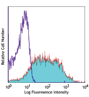
C57BL/6 mouse bone marrow cells were stained with B220 APC a... 
-
FITC anti-mouse CD117 (c-Kit)

C57BL/6 bone marrow cells were stained with CD45R/B220 PE an... 
-
PE anti-mouse CD117 (c-Kit)

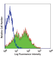
C57BL/6 bone marrow cells stained with 2B8 PE and CD11b FITC -
PE/Cyanine5 anti-mouse CD117 (c-Kit)
-
Purified anti-mouse CD117 (c-Kit)
C57BL/6 bone marrow cells stained with 2B8 PE and CD11b FITC -
PE/Cyanine7 anti-mouse CD117 (c-Kit)

C57BL/6 bone marrow cells stained with B220 FITC and 2B8 PE/... -
Alexa Fluor® 488 anti-mouse CD117 (c-Kit)

C57BL/6 mouse bone marrow cells stained with CD45R (RA3-6B2)... 
C57BL/6 mouse frozen testis section was fixed with 4% parafo... -
Alexa Fluor® 647 anti-mouse CD117 (c-Kit)

C57BL/6 mouse bone marrow cells stained with CD45R (RA3-6B2)... 
C57BL/6 mouse bone marrow cells stained with CD45R (RA3-6B2)... 
C57BL/6 mouse frozen testis section was fixed with 4% parafo... -
Pacific Blue™ anti-mouse CD117 (c-Kit)

C57BL/6 mouse bone marrow cells stained with CD45R (RA3-6B2)... 
C57BL/6 mouse bone marrow cells stained with CD45R (RA3-6B2)... -
PerCP/Cyanine5.5 anti-mouse CD117 (c-kit)

C57BL/6 mouse bone marrow cells stained with CD45R (RA3-6B2)... 
C57BL/6 mouse bone marrow cells stained with CD45R (RA3-6B2)... -
PerCP anti-mouse CD117 (c-kit)

C57BL/6 mouse bone marrow cells stained with CD45R (RA3-6B2)... 
C57BL/6 mouse bone marrow cells stained with CD45R (RA3-6B2)... -
APC/Cyanine7 anti-mouse CD117 (c-kit)

C57BL/6 mouse bone marrow cells were stained with CD11b FITC... -
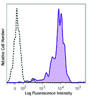
Brilliant Violet 421™ anti-mouse CD117 (c-Kit)


Confocal image of C57BL/6 mouse small intestine sample acqui... 
Confocal image of human lymph node sample acquired using the... -
Purified anti-mouse CD117 (c-Kit) (Maxpar® Ready)

Mouse bone marrow cells stained with 164Dy anti-Sca-1 (D7) a... -
PE/Dazzle™ 594 anti-mouse CD117 (c-Kit)

C57BL/6 mouse bone marrow cells were stained with CD11b FITC... 
-
Brilliant Violet 711™ anti-mouse CD117 (c-Kit)

C57BL/6 mouse bone marrow cells were stained with CD11b FITC... 
-
Alexa Fluor® 594 anti-mouse CD117 (c-Kit)

C57BL/6 mouse frozen testis section was fixed with 4% parafo... 
Paraformaldehyde-fixed (4%), 500 µm-thick mouse testis secti... -
APC/Fire™ 750 anti-mouse CD117 (c-Kit)

C57BL/6 mouse bone marrow cells were stained with CD11b PE a... 
-
Brilliant Violet 510™ anti-mouse CD117 (c-Kit)

C57BL/6 mouse bone marrow cells were stained with CD11b Alex... 
-
Brilliant Violet 785™ anti-mouse CD117 (c-Kit)

C57BL/6 mouse bone marrow cells were stained with CD11b Alex... 
-
TotalSeq™-A0012 anti-mouse CD117 (c-kit)
-
Brilliant Violet 605™ anti-mouse CD117 (c-Kit)

C57BL/6 mouse bone marrow cells were stained with CD11b FITC... -
Alexa Fluor® 700 anti-mouse CD117 (c-Kit)

C57BL/6 mouse bone marrow cells were stained with CD11b FITC... -
TotalSeq™-B0012 anti-mouse CD117 (c-Kit)
-
TotalSeq™-C0012 anti-mouse CD117 (c-Kit)
-
Spark NIR™ 685 anti-mouse CD117 (c-kit)

C57BL/6 mouse bone marrow cells were stained with CD11b FITC... -
Brilliant Violet 650™ anti-mouse CD117 (c-kit)

C57BL/6 mouse bone marrow cells were stained with CD11b FITC... -
Spark Red™ 718 anti-mouse CD117 (c-kit) (Flexi-Fluor™)


Follow Us