 Login / Register


- Clone
- RPA-T8 (See other available formats)
- Regulatory Status
- RUO
- Workshop
- IV T171
- Other Names
- T8, Leu2
- Isotype
- Mouse IgG1, κ
- Ave. Rating
- Submit a Review
- Product Citations
- publications

-

-

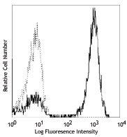
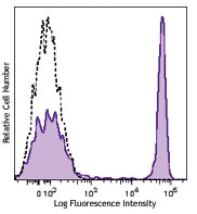
Human peripheral blood lymphocytes were stained with CD3 FITC and CD8 (clone RPA-T8) Brilliant Violet 421™ (top) or mouse IgG1, κ Brilliant Violet 421™ isotype control (bottom). -

Human peripheral blood mononuclear cells were fixed with 2% paraformaldehyde (PFA), then stained with 10 µg/ml anti-human CD8 (clone RPA-T8) Brilliant Violet 421™ (blue), 10 µg/ml of CD4 (clone RPA-T4) Alexa Fluor® 594 (cyan), and 10 µg/ml CD19 (clone HIB19) Alexa Fluor® 488 (green) for 30 minutes at room temperature. Nuclei were counterstained with DRAQ5 (red). The image was captured with a 40X objective.
| Cat # | Size | Price | Quantity Check Availability | Save | ||
|---|---|---|---|---|---|---|
| 301035 | 25 tests | 152€ | ||||
| 301036 | 100 tests | 305€ | ||||
CD8a is a 32-34 kD type I glycoprotein. It forms a homodimer (CD8a/a) or heterodimer (CD8a/b) with CD8b. CD8, also known as T8 and Leu2, is a member of the immunoglobulin superfamily found on the majority of thymocytes, a subset of peripheral blood T cells, and NK cells (which express almost exclusively CD8a homodimers). CD8 acts as a co-receptor with MHC class I-restricted T cell receptors in antigen recognition and T cell activation, and has been shown to play a role in thymic differentiation. Two domains in CD8a are important for function: the extracellular IgSF domain binds the α3 domain of MHC class I and the cytoplasmic CXCP motif binds the tyrosine kinase p56 Lck.
Product DetailsProduct Details
- Verified Reactivity
- Human, Cynomolgus, Rhesus
- Reported Reactivity
- Chimpanzee, Baboon, Pigtailed Macaque, Sooty Mangabey
- Antibody Type
- Monoclonal
- Host Species
- Mouse
- Formulation
- Phosphate-buffered solution, pH 7.2, containing 0.09% sodium azide and BSA (origin USA).
- Preparation
- The antibody was purified by affinity chromatography and conjugated with Brilliant Violet 421™ under optimal conditions.
- Concentration
- Lot-specific (to obtain lot-specific concentration and expiration, please enter the lot number in our Certificate of Analysis online tool.)
- Storage & Handling
- The antibody solution should be stored undiluted between 2°C and 8°C, and protected from prolonged exposure to light. Do not freeze.
- Application
-
FC - Quality tested
ICC - Verified - Recommended Usage
-
Each lot of this antibody is quality control tested by immunofluorescent staining with flow cytometric analysis. For flow cytometric staining, the suggested use of this reagent is 5 µL per million cells in 100 µL staining volume or 5 µL per 100 µL of whole blood. It is recommended that the reagent be titrated for optimal performance for each application.
Brilliant Violet 421™ excites at 405 nm and emits at 421 nm. The standard bandpass filter 450/50 nm is recommended for detection. Brilliant Violet 421™ is a trademark of Sirigen Group Ltd.
Learn more about Brilliant Violet™.
This product is subject to proprietary rights of Sirigen Inc. and is made and sold under license from Sirigen Inc. The purchase of this product conveys to the buyer a non-transferable right to use the purchased product for research purposes only. This product may not be resold or incorporated in any manner into another product for resale. Any use for therapeutics or diagnostics is strictly prohibited. This product is covered by U.S. Patent(s), pending patent applications and foreign equivalents. - Excitation Laser
-
Violet Laser (405 nm)
- Application Notes
-
The RPA-T8 antibody does not block the binding of HIT8a antibody to CD8a. Additional reported applications of this antibody (for the relevant formats) include: immunohistochemical staining of paraformaldehyde-fixed frozen sections3 and costimulation of T cell responses4. This clone was tested in-house and does not work on formalin fixed paraffin-embedded (FFPE) tissue. The Ultra-LEAF™ purified antibody (Endotoxin <0.1 EU/µg, Azide-Free, 0.2 µm filtered) is recommended for functional assays (Cat. Nos. 301073 & 301074).
-
Application References
(PubMed link indicates BioLegend citation) -
- Knapp W, et al. Eds. 1989. Leucocyte Typing IV. Oxford University Press. New York.
- Schlossman S, et al. Eds. 1995. Leucocyte Typing V. Oxford University Press. New York.
- Mack CL, et al. 2004. Pediatr. Res. 56:79. (IHC)
- Magidovich E, et al. 2007. P. Natl. Acad. Sci. USA 104:13022.
- Thakarl D, et al. 2008.J. immunol. 180:7431. PubMed
- Kmieciak M, et al. 2009. J. Transl. Med. 7:89. (FC) PubMed
- Thakral D, et al. 2008. J. Immunol. 180:7431. (FC) PubMed
- Yoshino N, et al. 2000. Exp. Anim. (Tokyo) 49:97. (FC)
- Rout N, et al. 2010. PLoS One 5:e9787. (FC)
- Stoeckius M, et al. 2017. Nat. Methods. 14:865. (PG)
- Product Citations
-
- RRID
-
AB_10898322 (BioLegend Cat. No. 301035)
AB_10960142 (BioLegend Cat. No. 301036)
Antigen Details
- Structure
- Ig superfamily, homodimer or heterodimer with CD8β, 32-34 kD
- Distribution
-
Majority of thymocytes, T cell subset, NK cells
- Function
- MHC class I co-receptor, thymic differentiation, T cell activation
- Ligand/Receptor
- MHC Class I molecules
- Cell Type
- Dendritic cells, NK cells, T cells, Thymocytes, Tregs
- Biology Area
- Immunology
- Molecular Family
- CD Molecules
- Antigen References
-
1. Barclay N, et al. 1993. The Leucocyte Antigen FactsBook. Academic Press Inc. San Diego.
- Gene ID
- 925 View all products for this Gene ID
- UniProt
- View information about CD8alpha on UniProt.org
Related Pages & Pathways
Pathways


Related FAQs
- What is the F/P ratio range of our BV421™ format antibody reagents?
-
It is lot-specific. On average it ranges between 2-4.
Other Formats
View All CD8a Reagents Request Custom ConjugationCustomers Also Purchased
Compare Data Across All Formats
This data display is provided for general comparisons between formats.
Your actual data may vary due to variations in samples, target cells, instruments and their settings, staining conditions, and other factors.
If you need assistance with selecting the best format contact our expert technical support team.
-
APC anti-human CD8a

Human peripheral blood lymphocytes stained with RPA-T8 APC -
APC/Cyanine7 anti-human CD8a

Human peripheral blood lymphocytes were stained with CD3 FIT... -
Biotin anti-human CD8a

Human peripheral blood lymphocytes stained with biotinylated... -
FITC anti-human CD8a

Human peripheral blood lymphocytes stained with RPA-T8 FITC -
PE anti-human CD8a

Human peripheral blood lymphocytes stained with RPA-T8 PE 
Human peripheral blood was stained with CD8a (clone RPA-T8) ... -
PE/Cyanine5 anti-human CD8a
-
PE/Cyanine7 anti-human CD8a

Human peripheral blood lymphocytes stained with RPA-T8 PE/Cy... -
Purified anti-human CD8a

Human peripheral blood lymphocytes stained with purified RPA... -
Alexa Fluor® 488 anti-human CD8a

Human peripheral blood lymphocytes stained with RPA-T8 Alexa... -
Alexa Fluor® 647 anti-human CD8a

Human peripheral blood lymphocytes stained with RPA-T8 Alexa... -
Pacific Blue™ anti-human CD8a

Human peripheral blood lymphocytes stained with RPA-T8 Pacif... -
Alexa Fluor® 700 anti-human CD8a

Human peripheral blood lymphocytes stained with RPA-T8 Alexa... -
PerCP anti-human CD8a

Human peripheral blood lymphocytes stained with RPA-T8 PerCP -
PerCP/Cyanine5.5 anti-human CD8a

Human peripheral blood lymphocytes were stained with CD3 APC... -
Brilliant Violet 421™ anti-human CD8a


Human peripheral blood mononuclear cells were fixed with 2% ... 
Human peripheral blood lymphocytes were stained with CD3 FIT... -
Brilliant Violet 570™ anti-human CD8a


Human peripheral blood lymphocytes were stained with CD3 and... -
Brilliant Violet 605™ anti-human CD8a

Human peripheral blood lymphocytes were stained with CD3 FIT... -
Brilliant Violet 650™ anti-human CD8a

Human peripheral blood lymphocytes were stained with CD3 FIT... -
Brilliant Violet 711™ anti-human CD8a

Human peripheral blood lymphocytes were stained with CD3 FIT... -
Brilliant Violet 785™ anti-human CD8a

Human peripheral blood lymphocytes were stained with CD3 FIT... -
Brilliant Violet 510™ anti-human CD8a

Human peripheral blood lymphocytes were stained with CD3 FIT... -
Purified anti-human CD8a (Maxpar® Ready)

Human PBMCs stained with 154Sm-anti-CD45 (HI30) and 146Nd-an... -
Alexa Fluor® 594 anti-human CD8a

Human peripheral mononuclear cells were fixed with 2% parafo... 
Human peripheral blood lymphocytes were stained with CD8 (cl... -
PE/Dazzle™ 594 anti-human CD8a

Human peripheral blood lymphocytes were stained with CD8 (cl... -
APC/Fire™ 750 anti-human CD8a

Human peripheral blood lymphocytes were stained with CD3 FIT... 
-
TotalSeq™-A0080 anti-human CD8a
-
TotalSeq™-B0080 anti-human CD8a
-
TotalSeq™-C0080 anti-human CD8a
-
Ultra-LEAF™ Purified anti-human CD8a

Human peripheral blood lymphocytes stained with LEAF™ purifi... -
Spark Violet™ 423 anti-human CD8a Antibody

Human peripheral blood lymphocytes were stained with anti-hu... -
Spark Red™ 718 anti-human CD8a

Human peripheral blood lymphocytes were stained with anti-hu... -
Spark PLUS UV™ 395 anti-human CD8a

Human peripheral blood lymphocytes were stained with anti-hu...


Follow Us