 Login / Register


Neuroprotection in the Parkinson’s Brain
 |
Protein aggregates are typically associated with pathogenic features in neurodegenerative diseases (e.g. α-Synuclein within Lewy bodies), but aggregation of the protein Parkin may actually be a marker of neuroprotection. Parkin primarily functions as an ubiquitin ligase that targets other proteins for degradation, and mutations in its gene cause autosomal recessive Parkinson’s disease (ARPD). A new study found that Parkin becomes insoluble during aging of a healthy human brain. Using novel antibodies that preferentially detect oxidized and aggregated forms of Parkin, researchers discovered that Parkin proteins protect neurons from oxidative stress and become aggregated in the process. These findings suggest that Parkin participates in both ubiquitin-proteasomal degradation and redox biology, two functions rarely possessed collectively by a single protein. Could these new insights inform the development of new treatments that restore Parkin function or relieve redox stress in patients with ARPD? |
| Parkin’s Role in Redox Biology Familial Parkinson’s disease (PD) is heritable and accounts for approximately 15% of all PD cases [1]. A mutated PRKN gene, which encodes the Parkin protein, is the most common cause of ARPD – a type of Familial Parkinson’s. Over 200 mutations of the PRKN gene have been identified, which can result in truncated forms of the protein and its functional loss [2]. Parkin is known to be involved in several aspects of neuronal biology, primarily ubiquitin-proteasomal degradation and mitochondrial maintenance. Loss of these functions from mutations, however, does not fully explain the specific death of dopaminergic neurons observed in ARPD. Previous work showing that Parkin deletion altered oxidative stress in animals led scientists to propose a new Parkin function: Parkin could play a role as a redox regulator, and loss of its anti-oxidant effects may contribute to neurodegeneration. |
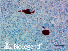 IHC staining with purified anti-Parkin antibody (clone A15165E) on formalin-fixed paraffin-embedded Parkinson’s disease substantia nigra tissue. BioLegend’s Ultra Streptavidin (USA) HRP Detection Kit (Multi-Species, AEC, Cat. No. 929801) was used for detection, followed by hematoxylin counterstaining. Our anti-Parkin (and PD-related) antibodies are currently discounted at 50% off; see list here. |
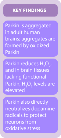 |
In their new study (Tokarew et al., now available in preprint on BioRxiv), Dr. Michael Schlossmacher’s lab utilized newly developed anti-Parkin antibodies to visualize the protein in brain tissues from humans ranging from 5 to 85 years of age. They found that Parkin was heavily aggregated in brain tissues from older humans, but could be disentangled into monomeric forms with reducing agents. This indicated that aggregated Parkin was oxidized, but what was causing this reaction? Since hydrogen peroxide (H2O2), a reactive oxygen species toxic to cells, was also elevated with age, the authors hypothesized that Parkin reduces H2O2 to relieve oxidative stress in young brains but becomes oxidized as a result. Aggregates of oxidized Parkin would then accumulate with age, leading to the phenotypes seen in older brain tissues. Parkin’s ability to reduce H2O2 was confirmed by in vitro assays, and analyses of samples from ARPD patients showed that H2O2 levels were indeed elevated in brain tissues deficient for normal Parkin. The researchers further demonstrated that rising levels of H2O2 in a dopamine toxicity model could be reduced by wildtype but not mutated Parkin, and that dopamine radicals could be directly neutralized by Parkin binding. Neutralization of these radicals caused the formation of neuromelanin, which were found to co-localize with Parkin in lysosomes. These results would explain Parkin’s specific protection of dopaminergic neurons and why this protection is lost with ARPD. |
| Advance Parkin Research with Aggregate-Preferring Clones The study by Tokarew et al. not only describes a novel role for Parkin in neuroprotection, but also sheds light on how its phenotypes evolve during aging in the brain. As new features of Parkin’s biology are uncovered, scientists will rely on the continual development of new tools for advancing research. BioLegend is committed to building partnerships with researchers so that we can deeply understand the needs of the field. Our collaboration with The Michael J. Fox Foundation and the Schlossmacher lab yielded the creation of anti-Parkin antibody clones A15165D and A15165E, both of which were used by Tokarew et al. to visualize Parkin in brain tissues. In their experiments, Tokarew et al. found both clones preferentially bound oxidized and aggregated forms of Parkin. If you’re interested in testing the application of these clones in your own studies, our anti-Parkin (and PD-related) antibodies are currently discounted at 50% off until May 31, 2020: |
| Specificity | Clone | Reactivity | Applications |
|---|---|---|---|
| Parkin | 5C3/Parkin | Human, Mouse, Rat | WB |
| Parkin (oxidized/aggregate preferring) | A15165D | Human | IHC-P, WB |
| Parkin (oxidized/aggregate preferring) | A15165E | Human | IHC-P |
(Promotional pricing cannot be combined with other discounts. Price reduction will be reflected during checkout. If you are a customer who orders via a distributor, please contact your distributor for eligibility.) Learn about other discounted antibodies for Parkinsons’ research Learn more about ‘Uncovering the Layers of Parkinson’s Disease’ by checking out our other blog post. Interested in receiving more related content? Sign up for our email updates. |
References:
|


Follow Us